Image guided thermal ablation of T1 renal cell cancer: A single centre experience.Image guided thermal ablation of T1 renal cell cancer: A single centre experience.Image guided thermal ablation of T1 renal cell cancer: A single centre experience.Image guided thermal ablation of T1 renal cell cancer: A single centre experience.

  • HOME
  • CONGRESS VENUE
  • SCIENTIFIC PROGRAMME
  • FACULTY
  • REGISTRATIONS
  • ABSTRACTS
  • EACCME® CREDITS
  • ITALIAN ECM ACCREDITATION
  • WHY ROME
  • ACCOMMODATION
  • DINNERS
  • GENERAL INFORMATION
  • SUPPORT
  • COI
  • E-Posters
Incidental findings we don’t want to miss on prostate multiparametric-MRI: a pictorial review
Settembre 9, 2023
Efficacy of MR-guided High Intensity Focused Ultrasound (MRgHIFU) in the treatment of painful bone metastases from prostate cancer
Settembre 9, 2023

Image guided thermal ablation of T1 renal cell cancer: A single centre experience.

Image guided thermal ablation of T1 renal cell cancer: A single centre experience.
J. Ryan (1), O. Lynch (2), B.B. McGuire (2), C.P Cantwell (1). 1. Department of Radiology, St. Vincent’s University Hospital, Dublin, Ireland. 2. Department of Urology, St. Vincent’s University Hospital, Dublin, Ireland.
Percutaneous image-guided tumour ablation (IGTA) with radiofrequency ablation (RFA), microwave ablation (MWA) and cryotherapy are curative local therapies for T1 renal cell carcinoma (RCC).
Although IGTA of T1 RCCs has been carried out for over 20 years, the long-term outcome data available on these patients has been limited to studies with small patient populations.
The aim of this study was to evaluate the oncological outcomes and survival times after ablation of T1 RCC in our unit over a 9-year period.
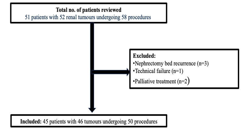
We retrospectively reviewed all patients with biopsy proven RCC who underwent IGTA of a T1 tumour between 1st March 2013 and 1st August 2022.
Figure 1. shows the total number of procedures reviewed with the inclusion/exclusion applied.
Ablation protocol - RFA was performed with a 2.5cm exposed 17 gauge cluster probe (Medtronic) and 12-minute impedance control ablation program.
MWA used a 14 gauge antenna.
The type of microwave program used depended on tumour location and size as well as consultant preference and manufacturer guidelines.
Cryotherapy used a 14 gauge applicator (Galil) and two freeze thaw cycles with two applicators placed 1 cm apart from each other and 1 cm from the tumour treatment margin.
Follow up protocol - Multiphasic renal CT or contrast-enhanced MRI at 3, 6, 9 and 12 months and then annually.
This protocol was followed within the confines of the limitation of radiological resources.
Data collected included: patient demographics, tumour size and histology, pre and post procedure creatinine, ablation technique, complications, charlston co-morbidity score, length of stay, oncological outcomes, disease free survival and overall survival.
Technical success was defined as completion of the treatment protocol.
Primary efficacy was defined as the absence of contrast enhancement within the ablated tumour on initial CT or MRI after the first ablative procedure.
Exlcusion-criteria
Figure 1. Flowchart of patient selection with inclusion criteria applied.
Patient demographics and tumour characteristics are summarized in Table 1.
Primary technical success was achieved in 100% of procedures (n=46/46).
Primary efficacy was achieved in 94% (43/46 tumours).   There was no statistically significant difference in mean pre-procedure creatinine and creatinine one month post-procedure [ 112 mm/l (52-792 mm/l) v 110 mm/l (53-748 mm/l), p = 0.75].
Median length of stay was one day (range 0-13 days).   Complications occurred after 8% of ablation procedures (4/50).
One patient had an intra-procedure pneumothorax that required a chest drain insertion.
The second patient had an intra-procedure haemothorax requiring a chest drain.
The third patient developed a perinephric collection at 3 months post ablation that resulted in ipsilateral mild hydronephrosis treated with image-guided drainage and antibiotic treatment as an inpatient on a second unscheduled admission.
One patient developed acute urinary retention post procedure.
Table 2 compares patient demographics, tumour size and periprocedural outcomes between RFA and MWA.
Primary efficacy was greater in the RFA group compared to the MWA group (p=0.02).
Larger median RCC diameter was associated with failed primary efficacy in the MWA group (3.8 cm vs 2.4 cm respectively, p= 0.045).
Overall survival and local tumour control within 9 years of IGTA are described in Figure 2 and 3, respectively.
Patient-characteristics
Table 1. Patient and tumour characteristics. Note: 1 patient underwent treatment for 2 separate RCC 8 years apart.
Comparison-MVA-v-RFA
Table 2. A comparison of microwave ablation (MWA) and radiofrequency ablation (RFA) group demographics, tumour characteristics and peri-procedural outcomes. Note: 1 patient underwent cryotherapy which is not included in the analysis.
Overall-Survival
Figure 2. Kaplan Meier graph demonstrating overall survival within 9 years after image guided tumour ablation.
In this study, overall survival at 1, 5 and 9 years was 97.4%, 66.7% and 40% respectively.
Local tumour control at 1, 5 and 9 years was 100%, 75% and 50% respectively.
In this study, primary efficacy was greater with RFA than MWA.
Primary efficacy failure in the MWA group was associated with greater median RCC diameters.
Local-tumour-control
Figure 3. Kaplan Meier graph demonstrating local tumour progression within 9 years after image guided tumour ablation.
Share
E-POSTER

  • [18F]-FACBC PET/CT Detection of Unusual and Usual Metastases in Prostate Cancer
  • 3D Magnetic Resonance Imaging–based Printed Models of Prostate before and after focal therapy

  • A Review of FDA Pharmacovigilance Database to Assess the Importance of Symptoms Associated with Gadolinium Exposure (SAGE)
  • Adjusted Skeletal Muscle Index (aSMI) correlates with outcome in metastatic Renal Cancer Cell (mRCC) patients (pts) treated with first line (1L) immunotherapy (IT)
  • ARTIFICIAL INTELLIGENCE IN FEMALE PELVIS MRI: CURRENT STATUS AND FUTURE PERSPECTIVES
  • Atypical leiomyoma or sarcoma? The great dilemma of the uterine myometrial tumors
  • Biopsy yields in different PSA density-PI-RADS score cohorts of biopsy-naïve patients with clinically-suspected prostate cancer
  • Birt-Hogg-Dubé syndrome: Review of radiological imaging , screening and follow-up recommendation.
  • Chronic painful cystitis and ureteric stricture following long term recreational Ketamine use
  • Chronic pelvic pain, can MRI play a role?
  • Comparison of a deep learning-accelerated T2-weighted turbo spin echo (TSE) sequence and a standard T2-weighted TSE in prostate MRI: reduced acquisition times and comparable image quality
  • Comparison of deep learning algorithms for prostate cancer detection​ and proposal of a new contrastive learning pre-training approach
  • Congenital Urachal Anomalies: Exploring the Spectrum of Imaging Findings
  • Contrast-enhanced MRI in women with endometrial cancer: dynamic versus single-phase acquisitions
  • Development of an interpretable CT radiomics model for the diagnosis of clear cell renal cell carcinoma in small solid renal masses
  • Diagnostic approach for intra-pelvic masses mimicking gynecologic masses
  • Diagnostic dilemmas and mimics of genito-urinary infections on imaging.
  • Diagnostic Performance of ADC vs ADCratio for the diagnosis of Prostate Cancer: Α Systematic review and Meta-analysis
  • Different faces of ovarian granulosa cell tumors: spectrum of MR imaging findings
  • Differentiation between an enlarged cyst and a hydronephrosis mimicking a large cyst: a critical checking point for guiding a treatment plan in PCKD patients with symptomatic or complicated cyst
  • Differentiation of endometriomas from ovarian hemorrhagic cysts on magnetic resonance imaging – a pictorial review
  • Diffusion-weighted imaging of the urinary tract – beyond tumors
  • Do MRI Structured Reports with FIGO classifications of Fibroids Contain Adequate Information for Clinical Decision Making?​
  • Does PSA density correlate with indicators of clinically significant prostate carcinoma?
  • Efficacy of MR-guided High Intensity Focused Ultrasound (MRgHIFU) in the treatment of painful bone metastases from prostate cancer
  • ENDOPROSTHESIS PLACEMENT IN PATIENTS WITH PREVIOUS SURGERY FOR OVARIAN CANCER COMPLICATED BY UROPERITONEUM
  • Evaluation of extraprostatic neoplastic extension through Multiparametric Magnetic Resonance of the Prostate.
  • Gastric-Type Adenocarcinoma of the Uterine Cervix: Role of Magnetic Resonance Imaging for Diagnosis and Management
  • Genitourinary fluoroscopic contrast examinations: what role do they have today?
  • Gynecological Masses: Tips for Determining their Site of Origin
  • Gynecological surgery’s complication: a radiological anatomical based approach to the diagnosis
  • High density flexible coil arrays for improved SNR in prostate MRI
  • High intensity focused ultrasound (HIFU) in the treatment of cancer of the prostate – Our experience
  • How embryology knowledge can help radiologists in the differential diagnosis of Wolffian duct (WD) related pathology
  • How to differentiate endometrial polyps from endometrial carcinomas on MRI: Tips and tricks
  • Image guided thermal ablation of T1 renal cell cancer: A single centre experience.
  • Image guided thermal ablation of T1 renal cell cancer: A single centre experience.
  • IMAGING FINDING IN RENAL CELL CARCINOMA – DIAGNOSIS AND STAGING
  • Imaging findings of primary prostatic lymphoma
  • Imaging in renal cell carcinoma – (a)typical findings
  • Impact of body composition on clinical outcomes in patients with metastatic hormone sensitive prostate cancer treated with androgen deprivation therapy plus second-generation antiandrogen.
  • Incidental detection on imaging in patients with a diagnosis of prostate cancer – lucky find or poisoned chalice?
  • Incidental detection on imaging performed during the work-up via MDTs of patients with kidney cancer – lucky find or poisoned chalice?
  • Incidental findings we don’t want to miss on prostate multiparametric-MRI: a pictorial review
  • Integrating clinical data with AI to optimise biopsy decisions after prostate MRI
  • Is it a leiomyoma? Challenging cases
  • MAP score and 2D Shear Wave ultrasound as adherent perinephric fat (APF) preoperative predictors in robot-assisted partial nephrectomy
  • mp-MRI appearance of primary and secondary prostate lymphomas
  • mpMRI may improve sensitivity and specificity of low dose short time 18F-PSMA 1007 imaging on a digital PET/CT scanner in low and medium risk prostate cancer patients after radical prostatectomy with biochemical recurrence at ultra-low PSA
  • MR guided Focal therapy – Spectrum of findings since the intervention day to long term surveillance.
  • MR imaging of abdominal pain in pregnant women
  • MR imaging of uterine fibroids – a pictorial review
  • MRI appearances of granulomatous prostatitis – our experience of 18 patients and pictorial review
  • MRI Characteristics of Corpus Luteum with Ovarian Oedema: Can both ovaries be affected?
  • MRI comparison of the two most actual classifications of the Mullerian anomalies
  • MRI in the characterization of renal masses
  • New transperineal ultrasound-guided biopsy for men in whom PSA is increasing after Miles’ operation
  • Novel CT-derived biomarkers for prediction of renal split function in preoperative nephrectomy planning
  • Patho-radiological correlation of clear cell renal cancer on double energy twim-beam abdominal computed tomography and biopsy samples
  • Pelvic and extra-pelvic endometriosis: a review of indications, techniques and MRI features.
  • Pelvic floor disorders: don’t forget men!
  • Pelvic venous congestion syndrome (PVCS) in women: a neglected pathology
  • Post-contrast acute kidney injury in patients with chronic kidney disease grade IV receiving iodinated contrast for elective computed tomography following new ESUR guidelines prophylaxis
  • PRACTICAL GUIDE TO PERFORM AND REPORT AN WB-DW-MRI STUDY FOR CARCINOMATOSIS OF GYNECOLOGICAL ORIGIN
  • Preoperative MRI assessment of hematometrocolpos with vaginal septum: added value of vaginal gel usage, reporting tips with surgical correlation.
  • Prospective evaluation of PI-RADS version 2.1 for clinically significant prostate cancer detection: Local performance at a University Hospital in Chile
  • Prostate MRI – are we good enough?
  • Radiomics for predicting extraprostatic extension of prostate cancer with MRI: a systematic review and meta-analysis
  • Rare presentations of testicular germ cell tumors: a pictorial multiparametric imaging review
  • Renal Ablation – Update of Current Guidelines and Impact on the Interventionalist
  • Renal Doppler Ultrasound for the prediction of non-diabetic kidney disease in diabetic patients
  • Renal Metastasis – all over the place
  • Role of MRI with diffusion-weighted imaging (DWI) in patients with uterine cervical cancer for intracavitary brachytherapy treatment alignment
  • Small renal masses management: a guidelines screenshot.
  • Soothing the headache of pregnancy and post-partum neurological complications: A pictorial review
  • The added value of capsular enhancement sign (CES) in the extra-prostatic extension (EPE) grading system in predicting prostate cancer with extracapsular extension
  • The Adequacy of Ultrasound Guided Native Renal Biopsy
  • The assessment of the Node Reporting and Data System (Node-RADS) in patients with cervical carcinoma
  • THE NEW 2019 BOSNIAK CLASSIFICATION OF COMPLICATED RENAL CYSTS. RADIOPATHOLOGICAL CORRELATION AND CRITICAL REVIEW.
  • The Prostate Imaging Quality Score (PI-QUAL): is inter-reader agreement a factor to consider?
  • The role of MRI in vulvar cancer staging according to the ESUR guidelines
  • The Role of Prophylactic Antibiotics in Transperineal Prostate Biopsy
  • The use of an AI software tool for prostate volume measurement on MRI
  • To determine the ability of an AI software tool to detect significant prostate cancer on MRI
  • To investigate the sonographic appearance of kidneys in farmers from a population-based study performed in a rural area of central-south Chile.
  • Untangling the Mystery: Radiological Insights into Female Pelvic Organ Torsion
  • Ureteral stone size measurements in non-enhanced CT (NECT) – interreader variability and prediction of spontaneous stone passage in different window settings
  • Using Low Energy Mono-Energetic CT Reconstruction to Improve Detection of  High-Risk Prostate Cancer
  • What is the best T1-weighted Dixon protocol for diagnosis of endometriosis?

Contact us


+39 06 52247328

FEDRA CONGRESSI S.A.S.
Viale dei SS. Pietro e Paolo, 21
00144 Roma
Tel +39.0652247328
Fax +39.065205625

www.fedracongressi.com
info@fedracongressi.com

  • HOME
  • CONGRESS VENUE
  • SCIENTIFIC PROGRAMME
  • FACULTY
  • REGISTRATIONS
  • ABSTRACTS
  • EACCME® CREDITS
  • ITALIAN ECM ACCREDITATION
  • WHY ROME
  • ACCOMMODATION
  • DINNERS
  • GENERAL INFORMATION
  • SUPPORT
  • COI
  • E-Posters
© 2023 Copyright by FEDRA CONGRESSI | All Rights Reserved |